
Diatoms are a widespread group and can be found in the oceans, in freshwater, in soils and on damp surfaces.
In Indonesia, during HackteriaLab 14, we tried to study the diversity of diatoms from the Kali-Code and see if diatoms can be used as a feasible bio-indicator of water pollution.
The first bottleneck was that the diatoms needed a slightly higher magnification than what the webcam microscope has in order to be observed clearly. We modified the webcam microscope by increasing the distance between the lens and the sensor to increase magnification. Later, back in the (Art)ScienceBLR lab, we rigged the webcam lens onto a DSLR to obtain clearer, magnified images of the diatoms.

The process we followed in Indonesia was also dependent on the use of a centrifuge. This made it a bit difficult for the process to be carried out in the field. If access to a centrifuge is difficult, samples can be swirled and left to settle in a test tube on the stand, for a crude separation during sedimentation. This method takes significantly longer, so it is recommended to use a centrifuge.

Using gaudilabs centrifuge to separate the diatoms. The diatoms form a white layer on top of the other algae.
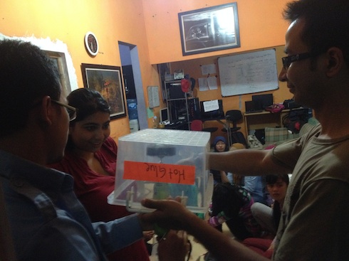

Centrifuging using the dremel at the workshop in Jogja
During the workshop we did with the interns of X-Code in Jogja, we also realized that because of the number of steps and complexity, this process would be better for a workshop which continues over a day or two, so that the information and process can be understood with clarity.


- Plastic tray
- Toothbrush with hard bristle preferably
- Drain cleaner (Anything containing sodium hypochlorite, Domex in India, Domestos Indonesia )
- Bigger eppendorf tubes, which can be used in a suitable centrifuge
- Test tubes and stand
- Pipet
- Plastic pipet/dropper
- A device which shows gps coordinates – smart phone in our case ( GPS test app)
- Microscpe ( A slighly modified webcam microscope is fine. We need a minimum magnification of around 300-400X.)
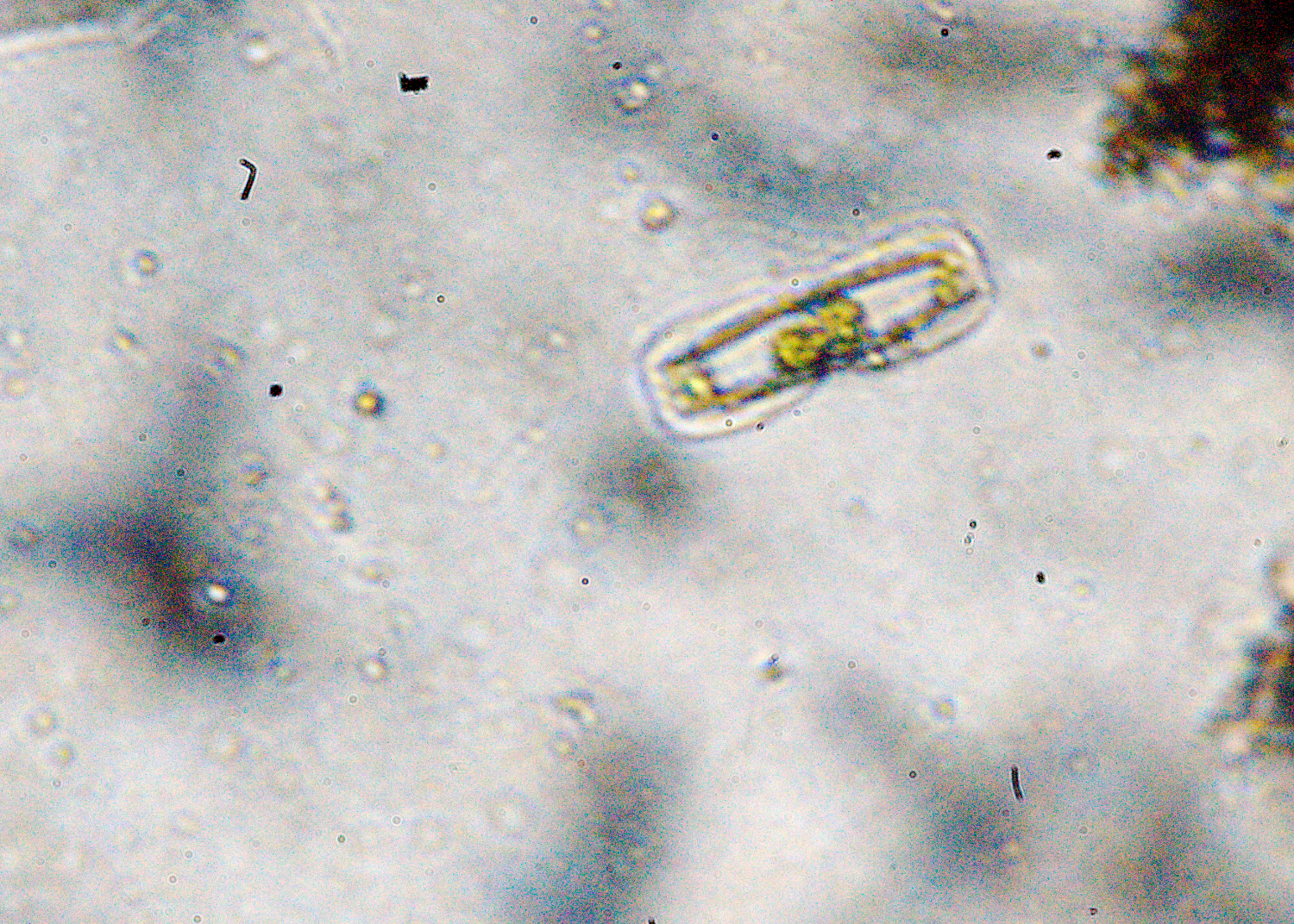
The diatoms are live here and some species are moving, which can make them hard to observe. If the concentration of the diatoms is high, this sample on the slide can be dried. After it has dried, The slide can be preserved using several methods. We used Akbars PVLG recipe and after mounting, sealed the slide with transparent nail polish.
8. Fill the tube with drain cleaner, and mix it through all the layers. Let this solution stand for half an hour, and agitate it 3 to 4 times while it sits.
11. After the final wash, the water can be thrown. The diatoms can be carefully removed from the white layer using a pipette and observed under the microscope. They can be mounted using Akbars PVLG recipe.
PVLG Receipe
Material:
100 mL distilled water
100 mL lactic acid
10 mL glycerol
16,6 g polyvinyl alcohol (PVA)
Preparation:
To prepare PVLG, add the polyvinyl alcohol (a dry powder) to the water and put in
60 degree oven until dissolves. Add lactic acid and glycerine and allow the solution
to set for 24 hours before first using. Specimens can be mounted directly in the
PVA solution, or the solution can be added to the sides of the cover slips that
were made with water, lactophenol, or melzer’s reagent. The PVA solution will
infiltrate the material in a day or two. Slides made with PVLG can be hardened by
heating at 40-75oC overnight. Immersion oil can be wiped from these hardened slides
without disturbing the specimen.
